&geometry(187x114) "ESANZ")
New ASE Guideline Helps Echocardiographers Recognise Cardiac Ultrasound Artifacts
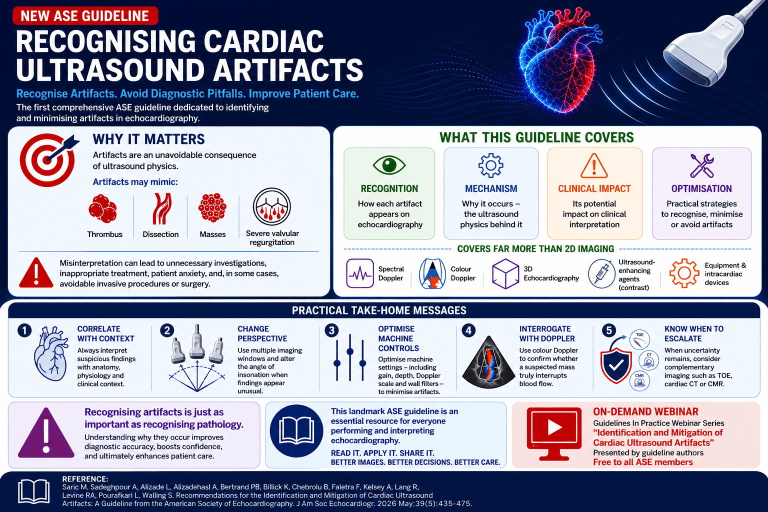
How confident are you in distinguishing an ultrasound artifact from true pathology?
The American Society of Echocardiography (ASE) has recently published its first comprehensive guideline dedicated entirely to cardiac ultrasound artifacts, Recommendations for the Identification and Mitigation of Cardiac Ultrasound Artifacts. This landmark document provides a practical, systematic approach to recognising, understanding, and mitigating artifacts encountered during echocardiographic imaging.
Artifacts are an inevitable consequence originating from the physical properties of ultrasound. While many are benign and easily recognised when their underlying mechanisms are understood; however, others can mimic significant pathology - including left atrial appendage thrombi, aortic dissections, intracardiac masses, or severe valvular regurgitation. Misinterpreting these findings can lead to unnecessary investigations, inappropriate treatment, and, in some cases, invasive procedures or surgery.
The guideline provides a practical, problem-solving framework by explaining:
- How each artifact appears on echocardiography
- Why it occurs, based on ultrasound physics
- Its potential clinical significance and diagnostic pitfalls
- Practical strategies to recognise, avoid, or minimise it during image acquisition and interpretation.
Importantly, the document extends beyond traditional 2D imaging to cover artifacts encountered in spectral Doppler, colour Doppler, 3D echocardiography, ultrasound-enhancing agents (contrast), as well as artifacts related to equipment and intracardiac devices.
Key Clinical Takeaways
Some of the most practical recommendations include:
- Correlate with context: Always interpret suspicious findings alongside clinical presentation, anatomy, and physiology.
- Change your perspective: Confirm unexpected findings by imaging from multiple acoustic windows and altering the angle of insonation.
- Optimise machine controls: Adjust gain, depth, Doppler scale, and wall filters to minimise artifacts before diagnosing pathology.
- Interrogate with Doppler: Use colour and spectral Doppler to assess flow patterns and to confirm whether a suspected intracardiac mass truly disrupts blood flow.
- Know when to escalate: When uncertainty remains, consider complementary modalities such as TEE, cardiac CT, or CMR.
For sonographers and echocardiographers, recognising artifacts is just as critical as identifying pathology. Understanding why artifacts occur improves diagnostic confidence, image quality, and ultimately patient care.
This landmark ASE guideline is a valuable educational resource for those performing or interpreting echocardiography and is well worth adding to your reading list.
To complement the guideline, the ASE and guideline chair Muhamed Saric, MD, PhD, FASE and co-chair Anita Sadeghpour, MD, FACC, FASE have hosted an on-demand webinar as part of the new Guidelines In Practice Webinar Series entitled “Identification and Mitigation of Cardiac Ultrasound Artifacts”. This webinar is available free to all ASE members.
Reference:
Saric M, Sadeghpour A, Alizade L, Alizadehasl A, Bertrand PB, Billick K, Chebrolu B, Faletra F, Kelsey A, Lang R, Levine RA, Pourafkari L, Walling S. Recommendations for the Identification and Mitigation of Cardiac Ultrasound Artifacts: A Guideline from the American Society of Echocardiography. J Am Soc Echocardiogr. 2026 May;39(5):435-475. doi: 10.1016/j.echo.2026.01.007.
)