&geometry(187x114) "ESANZ")

Educational Videos
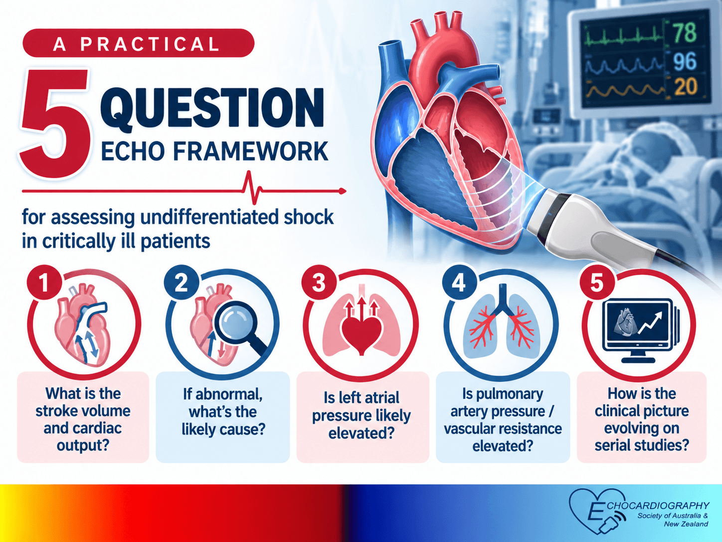 "5-Question Echo Approach to Undifferentiated Shock")
Prof Sam Orde interviews Prof Claire Colebourn and Dr Richard Fisher on their practical 5-question echo framework for assessing undifferentiated shock in critically ill patients.
 "Prosthetic Aortic Valves: SAVR vs. TAVR")
SAVR vs.TAVR - Do you know what makes them different?
Understand key haemodynamic, avoid measurement pitfalls, & improve the assessment of prosthetic aortic valve function with expert insights from Bianca Coelho.
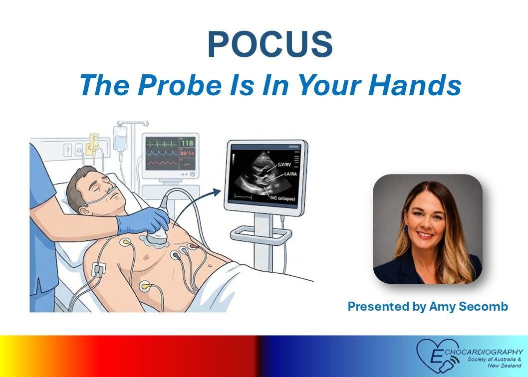 "Cardiac POCUS - The probe belongs in your hands!")
Join Amy Secomb, expert POCUS educator, who discusses the fundamentals of this valuable clinical tool.
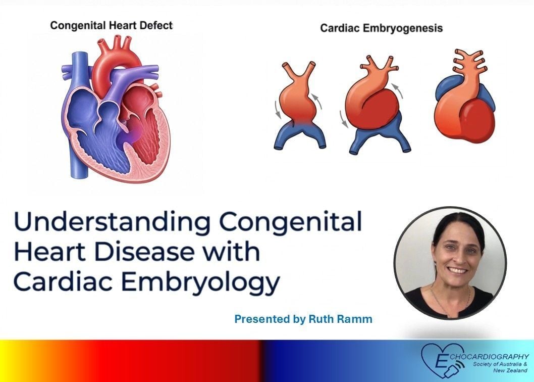 "Understanding cardiac embryology is the key to imaging congenital heart disease.")
Watch Ruth Ramm, widely acclaimed as an expert cardiac embryology educator, illuminate the complexities of the development of the human heart.
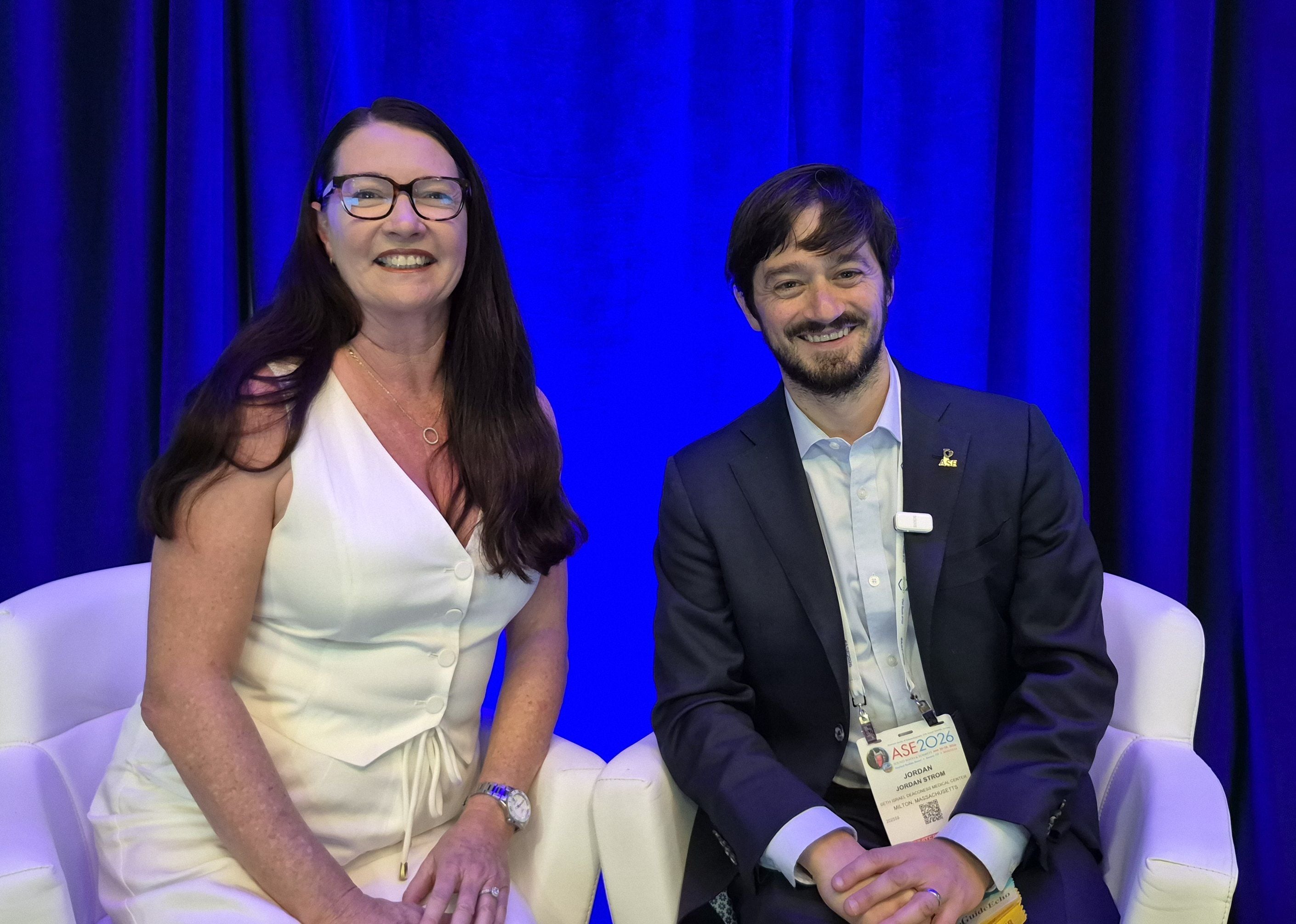 "ESANZ Meet the Echo Legends - Dr Jordan Strom")
Dr Julie Humphries interviews Echo Legend, Dr Jordan Strom
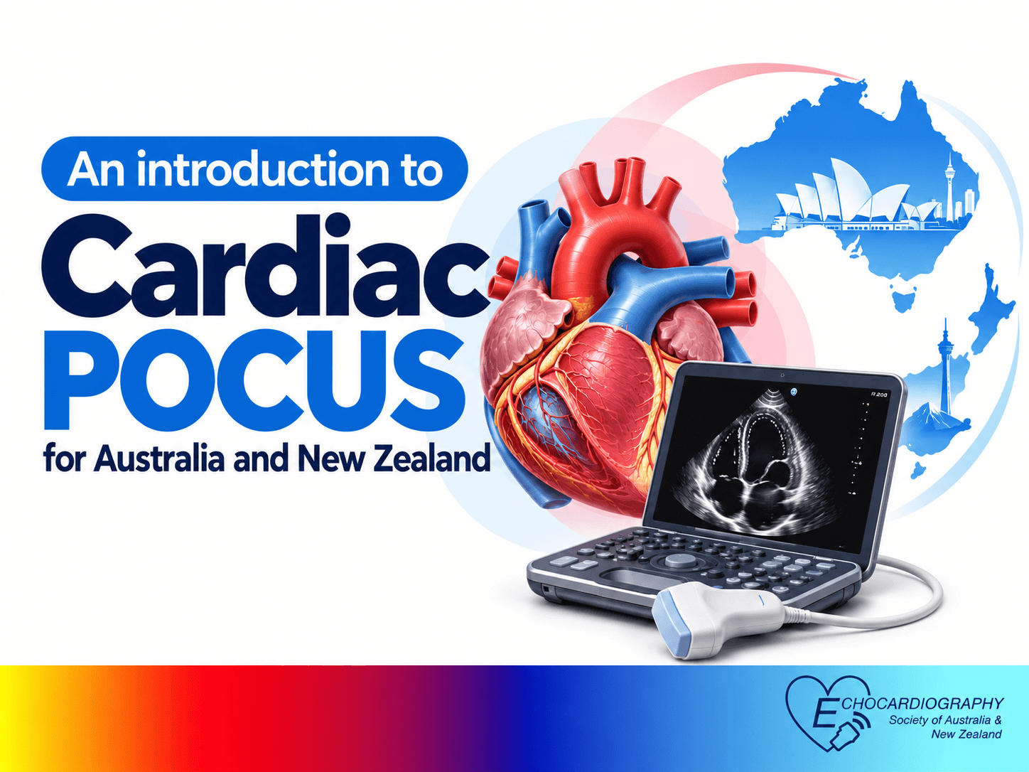 "Dr Drew Comeau gives an introduction to cardiac POCUS")
Join Dr Drew Comeau as he gives an overview of cardiac POCUS
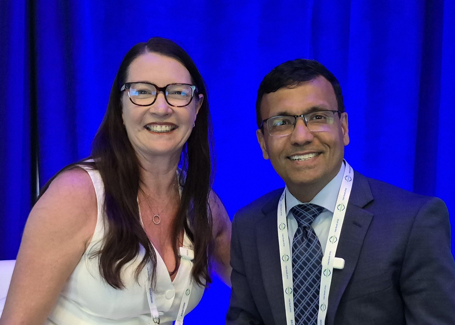 "ESANZ Meet the Echo Legends - Dr Partho Sengupta")
Dr Julie Humphries interviews Echo Legend, Dr Partho Sengupta.
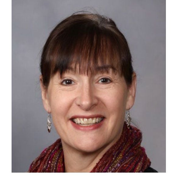 "ESANZ Meet the Echo Legends -Dr Merri Bremer")
Dr Julie Humphries interviews Echo Legend, Dr Merri Bremer, recipient of the 2026 Meritorious Service Award in honor of her distinguished career as an advanced cardiac sonographer and educator.
 "From ejection fraction to strain - the history of ventricular function assessment")
Join Professor Scalia for a nostalgic and insightful walk through the technologies that shaped, challenged, and refined how we measure left ventricular function.